Melanoma can spread in months or years; depth, ulceration, and lymph node findings shape the pace.
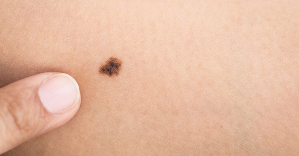
If a mole is changing, the first question is usually about time. That’s normal. The catch is that melanoma doesn’t run on one clock. One tumor can stay close to the surface for a long stretch. Another can grow downward early and reach a lymph node in a shorter window.
This article gives you a clean way to think about spread speed without making promises medicine can’t keep. You’ll see what “spread” means, what pathologists measure, what signs raise urgency, and what testing is commonly used when doctors need a clearer picture.
Why A Timeline Isn’t A Single Number
Spread is a chain. Cells must invade deeper skin, enter lymph or blood, survive travel, and then grow again elsewhere. Each step can move at its own pace. That’s why two melanomas with the same “age” on paper can behave in different ways.
There’s another snag: the clock often starts before you notice anything. A spot can blend in for a while, then change. So “I first saw it last month” may not match how long it’s been there.
What “Spread” Means In Melanoma
Doctors describe melanoma location in three main buckets. These buckets guide treatment and follow-up.
Local Disease
The cancer is only in the skin at the original site. It may be flat or raised, small or wide. The defining point is that melanoma cells aren’t found in nearby lymph nodes or far sites.
Regional Disease
The cancer has reached nearby lymph nodes or has formed small deposits in nearby skin or lymph channels. This is where risk rises and treatment options broaden. Stage III usually fits here.
Distant Disease
The cancer is found in organs or lymph nodes far from the original skin spot. This is stage IV on standard staging systems.
How Melanoma Moves From Skin To Nodes And Organs
Many melanomas start with side-to-side growth along the skin surface. Pathologists call this radial growth. In that phase, the spot can change shape and color without going far down.
When the tumor shifts into downward growth, called vertical growth, risk changes. Depth makes it easier for melanoma cells to reach lymph vessels and small blood vessels. Once cells enter those routes, spread can happen even if the original spot looks small.
The Depth Measurement That Drives Many Decisions
Your pathology report will usually list a number in millimeters called Breslow thickness. It measures how far melanoma cells extend into the skin. Depth is one of the strongest predictors of lymph node spread, and it steers choices like excision margins and whether node testing is offered.
Other Pathology Notes That Change Risk
Ulceration means the skin over the melanoma is broken on the microscope slide. At the same depth, an ulcerated tumor tends to act more aggressively than a non-ulcerated one. Reports may also mention cell division rate and whether cells are seen inside tiny vessels.
Types Of Melanoma And Typical Pace
Subtype and growth pattern can hint at pace, even before you see the final stage group.
- Superficial spreading melanoma often stays surface-first, then grows downward later.
- Nodular melanoma often grows downward early and may reach nodes sooner.
- Lentigo maligna melanoma often starts as a slow-growing patch on sun-exposed skin.
- Acral lentiginous melanoma appears on palms, soles, or nails and is often noticed late.
How Long Can Melanoma Take To Spread Through The Body
Skip the “deadline” idea. Think in bands tied to what the biopsy shows and what doctors find on exam.
- Melanoma in situ is limited to the top skin layer. It can widen across the surface, yet it hasn’t entered deeper routes that lead to nodes or organs.
- Thin invasive melanoma has entered deeper skin but remains shallow. Many cases are treated before they ever reach a node.
- Thicker or ulcerated melanoma has higher odds of reaching a node, and that can happen within months in some cases.
- Node-positive or distant melanoma is already beyond the original site. Timing then depends on where disease is found and how it responds to treatment.
Stage labels are built from these facts. For the official stage definitions doctors use, see the American Cancer Society stage breakdown for melanoma.
| Clue | What It Can Suggest | Common Next Step |
|---|---|---|
| In situ (top layer only) | No invasion into deeper skin yet | Excision with margins; usually no node test |
| Under 1 mm depth | Lower odds of node spread, not zero | Wide excision; node test in selected cases |
| 1–2 mm depth | Rising chance of regional spread | Sentinel lymph node biopsy often offered |
| 2–4 mm depth | Higher odds of node involvement | Node testing is common; imaging may be added |
| Over 4 mm depth | Higher chance of spread beyond skin | Node testing plus imaging, based on the full picture |
| Ulceration present | Higher-risk behavior at the same depth | Stage group shifts; follow-up may be tighter |
| Positive sentinel lymph node | Regional spread is present | Stage III plan; systemic therapy may be offered |
| In-transit or satellite lesions | Cells have moved through nearby lymph channels | Stage III workup; imaging and systemic therapy may be used |
| Distant metastasis found | Cells have reached far sites | Stage IV plan with systemic therapy and targeted imaging |
How Long Does It Take For Melanoma To Spread?
Here’s the practical takeaway: spread risk is tied more to depth and lymph node status than to calendar time. A thin melanoma can be removed before it reaches a node. A thicker melanoma can reach a node in months. That’s why a biopsy and a complete pathology report matter so much.
Population data shows the same pattern. Using SEER stage groupings, the American Cancer Society reports five-year relative survival above 99% for localized melanoma, 76% for regional melanoma, and 35% for distant melanoma, based on U.S. cases diagnosed from 2015 to 2021 in its survival rates by stage page. Those are group averages, not personal predictions, yet they show why early evaluation can change outcomes.
Treatment choices are stage-based. The National Cancer Institute’s Melanoma Treatment (PDQ®) summary shows how care changes from early disease to unresectable stage III and stage IV disease.
Signs That Raise Urgency
Melanoma can spread without symptoms, so skin change is often the first signal. The CDC’s melanoma warning signs list uses the “ABCDE” pattern that many clinicians teach.
ABCDE Skin Changes
- Asymmetry: one half doesn’t match the other.
- Border: edges look jagged, blurred, or uneven.
- Color: mixed colors in one spot, or a new dark area.
- Diameter: growth in size, or a small lesion changing fast.
- Evolving: any shift in size, shape, color, bleeding, or itch.
Clues Near The Original Spot
New small bumps near the lesion, or a firm cord under the skin, can happen when cells move through local lymph channels. Swelling in nearby lymph nodes can be painless, so it’s easy to miss unless you check.
Symptoms Away From The Skin
Persistent cough, ongoing headaches, bone pain, or unplanned weight loss can have many causes. If melanoma is already diagnosed, new symptoms like these should lead to a call to your care team.
How Doctors Check If Melanoma Has Spread
The diagnostic path is usually stepwise. Doctors start with proof, then build staging from what they find.
Skin Biopsy And Pathology Report
A biopsy confirms melanoma and gives the first staging clues: depth, ulceration, and margin status. Ask for a copy of the pathology report. It’s the document every specialist will want.
Wide Local Excision
After diagnosis, surgeons remove the melanoma site with a rim of normal-looking skin. Margin size is tied to depth and location. This treats the primary site and lowers the chance of local return.
Sentinel Lymph Node Biopsy
For tumors above certain risk thresholds, doctors may offer a sentinel lymph node biopsy. A tracer maps the first draining node or nodes. Those nodes are removed and checked for melanoma cells. A positive result often moves staging into stage III territory.
Imaging And Blood Work
Scans aren’t routine for every early melanoma. They’re more common when stage, exam findings, or symptoms suggest a higher chance of spread. Imaging choice depends on what doctors are trying to rule out and which body sites are in question.
| Test | When It’s Used | What It Checks |
|---|---|---|
| Skin biopsy | First step for a suspicious lesion | Diagnosis, depth, ulceration, margins |
| Pathology review | After biopsy | Cell division rate and other high-risk notes |
| Sentinel node biopsy | Often when depth is near or above 1 mm | Microscopic spread to first draining nodes |
| Node ultrasound | When nodes feel enlarged, or in follow-up | Node size and structure |
| CT / PET / MRI | When stage or symptoms raise concern | Distant spread to organs or brain |
| LDH blood test | Often in advanced disease workups | A marker used in prognosis in some settings |
What To Do If You’ve Spotted A Suspicious Change
A clear next step can calm the mind and cut delay. Here’s a plan that fits most situations.
Do A Two-Minute Record
- Take sharp photos in good light. Add a ruler or coin for scale.
- Write down the date you noticed the change and what changed.
- Check nearby skin for new bumps. Feel for swollen nodes in the nearest basin.
Book The Right Visit
A dermatologist is a good starting point. If you can’t get in soon and the lesion is bleeding or changing fast, urgent care or a primary doctor visit is still smart. Ask for a full skin exam and a biopsy plan.
Bring These Questions
- What is the Breslow thickness and is there ulceration?
- Do you recommend sentinel lymph node biopsy for my case?
- What follow-up schedule fits my stage, and what changes should trigger a call?
- If staging is III or IV, what systemic treatments match the tumor’s markers?
Act On The Pathology Report
If melanoma is confirmed, ask when the wide excision is scheduled and when final staging will be ready. Keep your pathology report handy so new clinicians can move faster.
How This Article Was Built
This article uses staging definitions and public agency guidance listed below to frame spread timing without guessing a single “average” clock.
References & Sources
- American Cancer Society.“Stages of Melanoma Skin Cancer.”Stage definitions tied to depth, lymph nodes, and distant spread.
- American Cancer Society.“Survival Rates for Melanoma Skin Cancer, by Stage.”SEER stage survival ranges used to show how outcomes differ by stage.
- Centers for Disease Control and Prevention (CDC).“Symptoms of Skin Cancer.”ABCDE warning signs and other skin changes that should prompt evaluation.
- National Cancer Institute (NCI).“Melanoma Treatment (PDQ®).”Stage-based treatment options, including care for unresectable stage III and stage IV disease.

Mo Maruf
I created WellFizz to bridge the gap between vague wellness advice and actionable solutions. My mission is simple: to decode the research and give you practical tools you can actually use.
Beyond the data, I am a passionate traveler. I believe that stepping away from the screen to explore new environments is essential for mental clarity and physical vitality.